
Blood Vessel Stenosis
Imaging via H&E Histology
While the histology can be measured live on a microscope, it is easier to stitch an image from several high resolution photos or take one photo at low resolution.

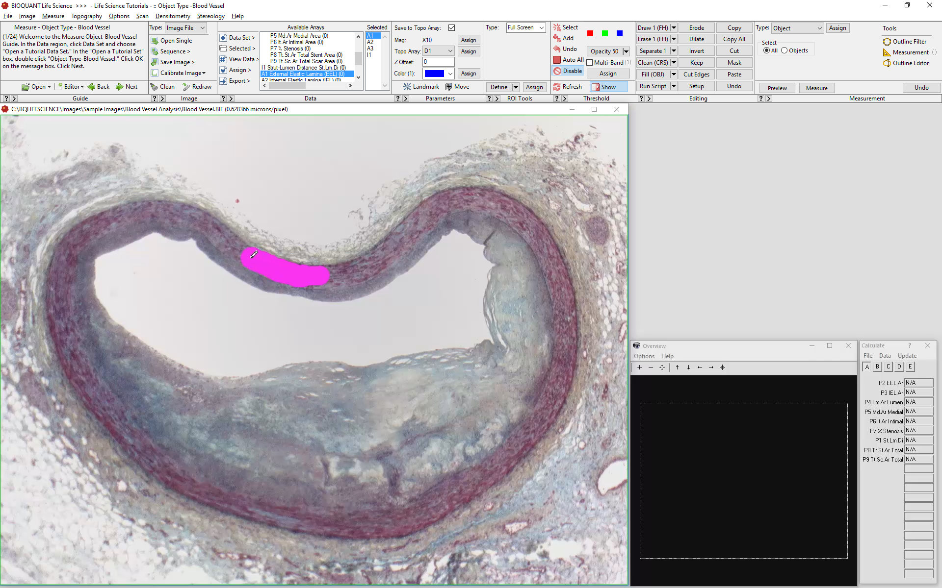
The EEL is Defined and Edited
Using BIOQUANTs Editing Tools, the histologist defines the EEL boundaries. Once defined, BIOQUANT will use the defined threshold to automatically measure the EEL and IEL.
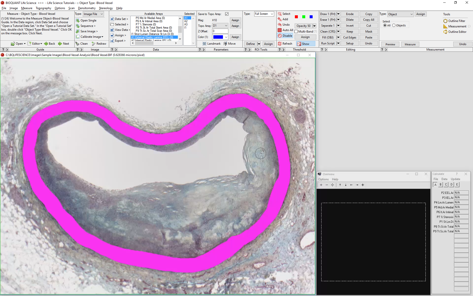
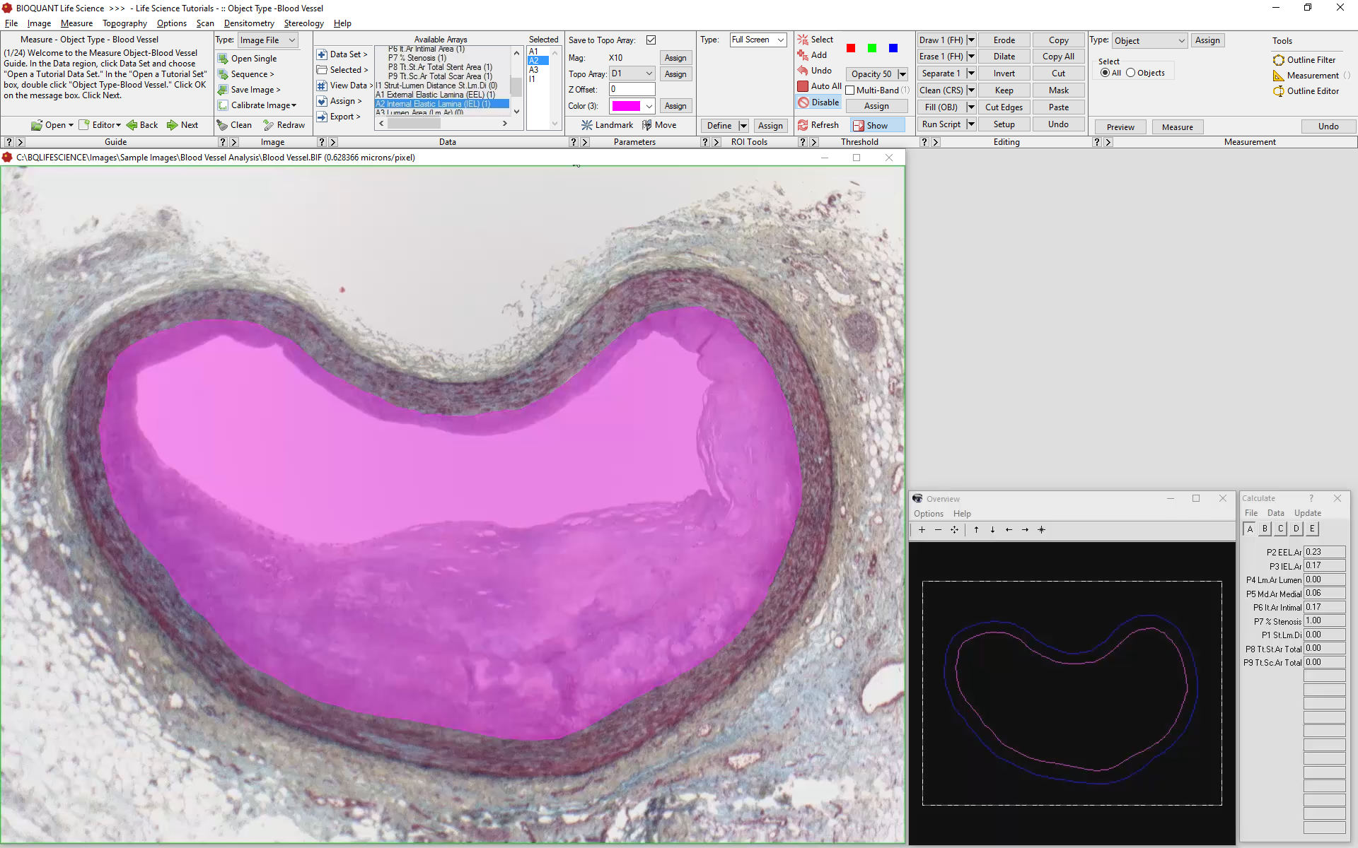
Measure the EEL
Once defined, Measure the EEL.

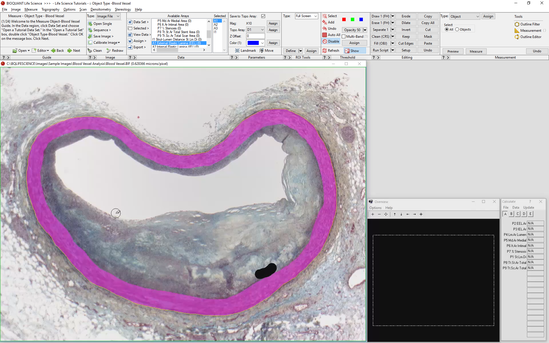
Using the Existing Threshold, Limit to the IEL
Measure the IEL
Measure the IEL based on the modified threshold.

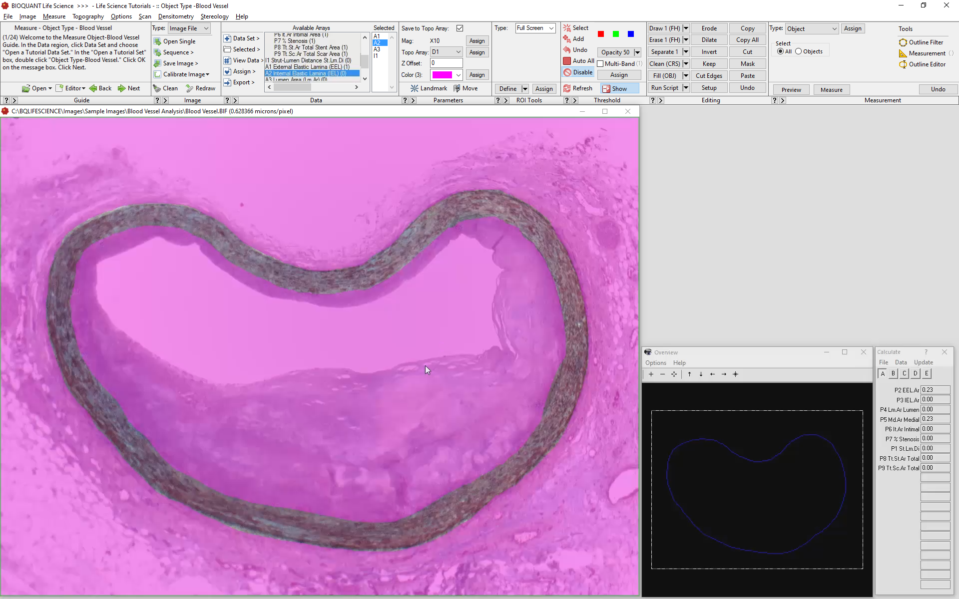
Threshold and Limit the Lumen Area
The lumen has enough contract from surrounding tissue to be automatically measured. Threshold the Lumen, then, using BIOQUANTs Editing Tools, Keep the Lumen.
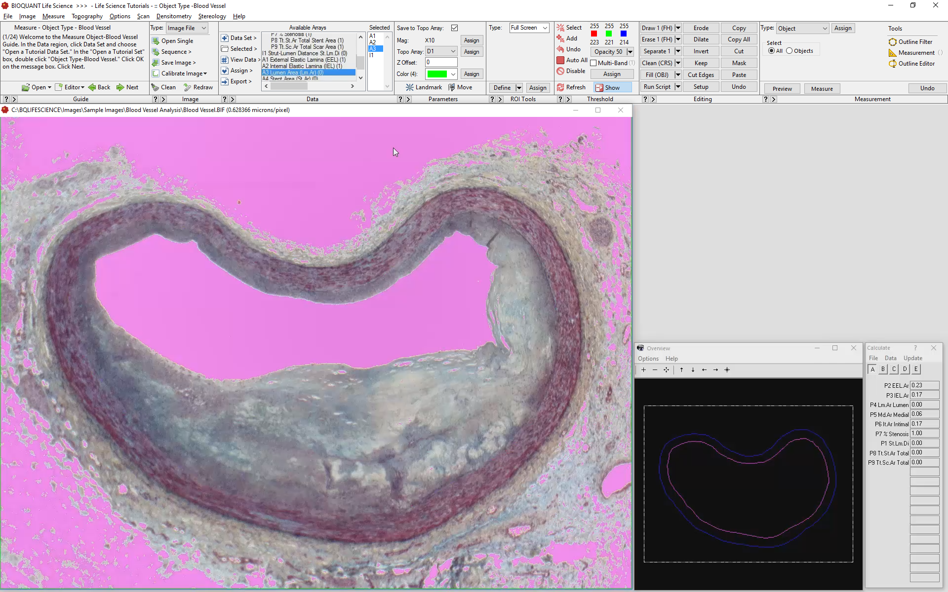

Measure the Lumen
BIOQUANT will automatically collect the Lumen data and perform calculations using the updated information.

Stenosis and Stent Performance
Once the primary data are collected, the software internally processes the data to produce a final report.
Images with or without schematic markup are saved to compliment the quantitative data.
Computed Data
Stent to Lumen Distance
External Elastic Lamina Area
Internal Elastic Lamina Area
Lumen Area
Medial Area
Intimal Area
% Stenosis
Total Stent Area
Total Scar Area